anti-Caspase-1 (p20) (mouse), mAb (Casper-1)
[Interleukin-1β Convertase; IL-1BC; Interleukin-1β-converting Enzyme; ICE]
AG-20B-0042-C100 100 μg
Clone Casper-1
Source/Host Purified from concentrated hybridoma tissue culture supernatant.
Isotype Mouse IgG1
Immunogen Recombinant mouse caspase-1.
Handling / Storage
Shipping BLUE ICE
Short Term Storage +4°C
Long Term Storage -20°C
After opening, prepare aliquots and store at -20°C. Avoid freeze/thaw cycles.
Use / Stability
Stable for at least 1 year after receipt when stored at -20°C.
MSDS available at www.adipogen.com or upon request.
Product Specifications
Specificity Recognizes endogenous full-length and activated (p20 fragment) mouse caspase-1.
Species Crossreactivity Mouse
Application Western Blot (see online protocol): (1μg/ml) (no need to precipitate the cell supernatant
for the detection of caspase-1 (mouse) upon inflammasome activation)
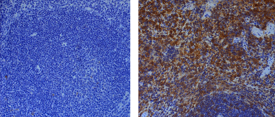
Immunohistochemistry: (1:500; paraffin sections)
Immunoprecipitation: (1:200)
Purity ≥95% (SDS-PAGE)
Formulation Liquid. In PBS containing 10% glycerol and 0.02% sodium azide.
Concentration 1mg/ml
Product Description
Caspase-1 is the best-described inflammatory caspase. It processes the cytokines interleukin-1β (IL-1β) and
IL-18 and induces pyroptotic cell death. Caspase-1 is activated by multiprotein complexes called Inflammasomes
in response to numerous stimuli that are detected through distinct inflammasomes. NLRC4 responds to cytosolic
flagellin, murine NLRP1b responds to anthrax lethal toxin, AIM2 responds to cytosolic DNA and NLRP3 responds
to a variety of agonists including crystals.

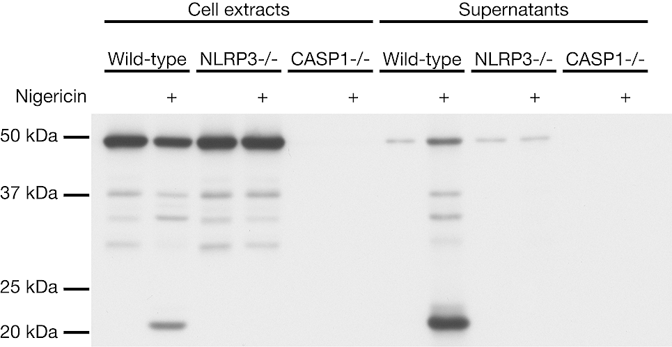
Figure 1: Mouse caspase-1 (p20) is detected by immunoblotting using anti-Caspase-1 (p20) (mouse), mAb
(Casper-1) (Prod. No. AG-20B-0042). Method: Caspase-1 was analyzed by Western blot in cell extracts and
supernatants of differentiated bone marrow-derived dendritic cells (BMDCs) from wild-type, NLRP3-/- and caspase-
1-/- mice activated or not by 5 μM Nigericin (Prod. No. AG-CN2-0020) for 30 min. Cell extracts and supernatants
were separated by SDS-PAGE under reducing conditions, transferred to nitrocellulose and incubated with anti-
Caspase-1 (p20) (mouse), mAb (Casper-1) (1μg/ml). Proteins were visualized by a chemiluminescence detection
system.


 010-64814275
 當前位置:
當前位置:


 010-64814275
 010-64814275
 2850669802