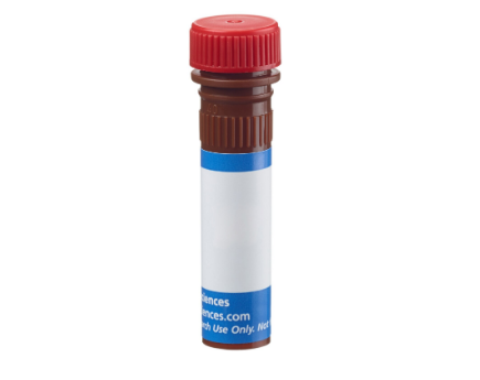
BrandBD Pharmingen™
Alternative NameIFNG; Interferon-gamma; IFG; IFI; Type II interferon
Vol. Per Test20 µl
IsotypeMouse BALB/c IgG1, κ
ReactivityRhesus, Cynomolgus, Baboon (QC Testing) Human (Tested in Development)
ApplicationIntracellular staining (flow cytometry) (Routinely Tested)
ImmunogenHuman IFN-γ from supernatants of S. aureus-stimulated PBMC
Storage BufferAqueous buffered solution containing BSA and ≤0.09% sodium azide.
Description
The 4S.B3 monoclonal antibody specifically binds to interferon-γ (IFN-γ). The immunogen used to generate this hybridoma was partially purified human IFN-γ obtained from supernatants of human PBMC stimulated with Staphylococcus aureus. Interferon-γ (IFN-γ) is a potent multifunctional cytokine that is produced by several activated cell types including NK, NKT, CD4+TCRαβ+, CD8+TCRαβ+, and TCRγδ+ T cells. IFN-γ exerts its biological effects through specific binding to the high-affinity IFN-γ Receptor Complex comprised of IFN-γRα (CD119) and IFN-γRβ subunits. In addition to its antiviral effects, IFN-γ upregulates a number of lymphoid cell functions including the antimicrobial and antitumor responses of macrophages, NK cells, and neutrophils. In addition, IFN-γ can exert strong regulatory influences on the proliferation, differentiation, and effector responses of B cell and T cell subsets. These influences can involve IFN-γ's capacity to boost MHC class I and II expression by antigen-presenting cells as well as to direct effects on B cells and T cells themselves. Human IFN-γ is a 14-18 kDa glycoprotein containing 143 amino acid residues.
Clone 4S.B3 also cross-reacts with a cytoplasmic component of peripheral blood CD3+ lymphocytes of baboon, and both rhesus and cynomolgus macaque monkeys following five-hour treatment with phorbol myristic acetate (PMA) and Ca++ ionophore (A23187) in the presence of monensin. The staining pattern of 4S.B3 in CD3+ cells is similar to that observed with peripheral blood T lymphocytes from normal human donors. This reagent is useful for intracellular immunofluorescent staining for flow cytometric analysis to identify and enumerate IFN-γ + cells within a mixed cell population.
FORMAT

FormatPE
Excitation SourceBlue 488 nm,Green 532 nm,Yellow/Green 561 nm
Excitation Max496 nm
Emission Max578 nm
R-phycoerythrin (PE) is an accessory photosynthetic pigment found in red algae. It exists in vitro as a 240-kDa protein with 23 phycoerythrobilin chromophores per molecule. This makes PE one of the brightest fluorochromes for flow cytometry applications, but its photobleaching properties make it unsuitable for fluorescence microscopy.

 010-64814275
 當前位置:
當前位置:


 010-64814275
 010-64814275
 2850669802